
Fig. S5
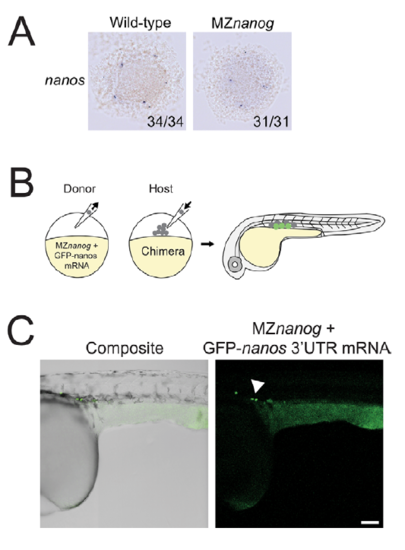
MZnanog germ cells are specified and migrate correctly when transplanted into wild-type host embryos. A. In situ hybridization for nanos expression at sphere stage in wildtype and MZnanog embryos. B. A diagram of transplantation of cells from MZnanog embryos injected with 50 pg GFP-nanos1 3’UTR mRNA (“GFP-nanos”, Köprunner et al., 2001) into a wild-type host embryo, with green cells in the host embryo indicating expression of the mRNA in transplanted cells at 30 hpf. C. Approximately 20 cells were transplanted from donor injected embryos into uninjected wild-type host embryos. At 30 hpf, embryos were anaesthetized, mounted, imaged, and processed as in Figure 6D (n=6). Germ cells are indicated with an arrowhead. Scale bar indicates 100 microns. |