
Fig. 2
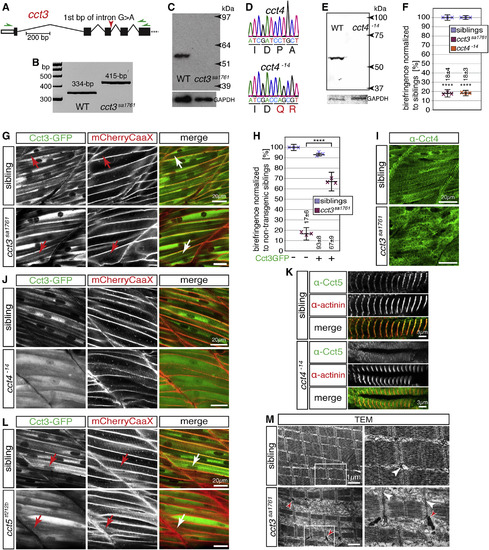
Loss of TRiC Subunits Causes Lack of Functional TRiC at Sarcomeric Z-disks (A) cct3sa1761 harbors an essential splice site mutation in cct3 (red arrow). (B) RT-PCR using oligonucleotides targeting regions marked by green arrows in (A) generated a 334-bp amplicon with WT embryos and a single 415-bp amplicon with cct3sa1761 homozygotes. Amplicons were identified by sequencing. (C) Western blot analysis using antibodies against human CCT3 revealed epitope loss in cct3sa1761 homozygotes (GAPDH served as a loading control). (D) CRISPR/Cas9-mediated deletion of 14 bp from exon 5 of cct4 causes a frameshift evoking premature stop codons in cct4−14. (E) Western blot analysis using antibodies against CCT4 revealed epitope loss in cct4−14 homozygotes (GAPDH served as a loading control). (F) At 3 dpf, the birefringence was significantly reduced in cct3sa1761 and cct4−14 mutants compared with siblings. Data are mean ± SEM; ∗∗∗∗p < 0.0001 by Student’s t test; n = 3. (G) GFP fluorescence of Tg(cry:mCherry,-600unc:cct3GFP) localized to sarcomeric Z-disks as identified by co-localization with mCherryCaaX-positive t-tubules introduced by Tg(acta1:mCherryCaaX) in siblings and cct3sa1761 homozygotes (arrows) (n = 5 per genotype). (H) Expression of the Cct3-GFP fusion protein ameliorated the birefringence of cct3sa1761 homozygotes with high significance. Data are mean ± SEM; ∗∗∗∗p < 0.0001 by one-way ANOVA with Tukey’s post hoc test; n = 3. (I) The striated pattern obtained with antibodies against Cct4 in siblings was severely compromised in cct3sa1761 (n = 5 per genotype). (J) Compared with siblings, localization of Cct3-GFP to Z-disks is severely compromised in cct4−14 homozygotes transgenic for Tg(cry:mCherry,-600unc:cct3GFP) and Tg(acta1:mCherryCaaX) (n = 4 per genotype). (K) Immunohistochemistry detected the localization of Cct5 at actinin-positive Z-disks in siblings but not in cct4−14 homozygotes (n = 3 per genotype). (L) In cct5tf212b homozygotes and siblings that harbor Tg(cry:mCherry,-600unc:cct3GFP) and Tg(acta1:mCherryCaaX), GFP-tagged Cct3 localized to Z-disks (arrows) (n = 3 per genotype). (M) Representative TEM micrographs of cct3sa1761 present a reduced amount of myofibrils (n = 3 per genotype). Importantly, although the sarcomeres appear organized, electron-dense rods were detected at Z-disks in cct3sa1761 (red arrowheads). White arrowheads point to t-tubules at Z-disks. Boxed areas are shown at higher magnification. See also Figures S4, S5, and S7. |
| Genes: | |
|---|---|
| Antibodies: | |
| Fish: | |
| Anatomical Terms: | |
| Stage: | Protruding-mouth |
| Fish: | |
|---|---|
| Observed In: | |
| Stage: | Protruding-mouth |