
FIGURE
Fig. S7
Fig. S7
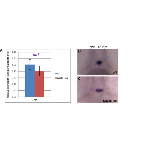
Kctd15 mutants show gh RNA levels similar to wild type. gh levels were examined by qPCR (A) and in situ hybridization (B) in WT and mutant embryos at 48 hpf. While there is a general trend towards lower gh levels, this difference is not significant. In ~60% of embryos, the staining pattern of gh transcripts appears more sparse (in fewer cells), when compared to the rosette pattern observed in WT. |
Expression Data
Expression Detail
Antibody Labeling
Phenotype Data
Phenotype Detail
Acknowledgments
This image is the copyrighted work of the attributed author or publisher, and
ZFIN has permission only to display this image to its users.
Additional permissions should be obtained from the applicable author or publisher of the image.
Full text @ PLoS One