Fig. s2
- ID
- ZDB-FIG-180417-16
- Publication
- Sylvester et al., 2017 - Population-scale organization of cerebellar granule neuron signaling during a visuomotor behavior
- Other Figures
- All Figure Page
- Back to All Figure Page
|
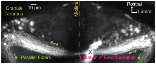
Targeting granule neurons for antidromic activation. To determine how changes in granule cell firing were coupled to changes in the somatic calcium concentration, we sought to identify a location for electrical stimulation of granule cell axons that would enable antidromic activation of granule neurons in the absence of direct depolarization to the soma or secondary input from mossy fibers or other cerebellar neuronal classes. Here we show that targeting of the caudal molecular layer in one half of the cerebellum allows selective access to many of the granule cells with somata in the other half of the cerebellum. To do so, we targeted caudal lobe commissure parallel fibers1 for electroporation with 3 kD Texas Red Dextran (D-3329, Life Technologies; 10 mM in distilled water; 7 dpf nacre). Electroporation via a fine glass capillary (1 second long train of 2 msec 70 V pulses delivered at 200 Hz) resulted in immediate loading of cells and tissue near the poration site (“X”). Over a course of ten minutes, dye diffused in parallel fibers away from the poration site to label granule cell somata located on either side of the midline within the eminentia granularis and lateral portions of the IGL. Some parallel fibers were also seen to exit the cerebellum on both sides (green arrows) and terminate in rhombomeres 1 and 2 (not shown). For the antidromic activation experiments found within the main text, depolarizing electrical pulses were therefore delivered at locations similar to the location of the electroporation shown here, thereby minimizing activation of a larger portion of the cerebellar circuit and ensuring that changes in somatic fluorescence intensity were directly driven by changes in granule cell firing. |

