
Fig. 3
- ID
- ZDB-FIG-180403-20
- Publication
- Roh-Johnson et al., 2017 - Macrophage-Dependent Cytoplasmic Transfer during Melanoma Invasion In Vivo
- Other Figures
- All Figure Page
- Back to All Figure Page
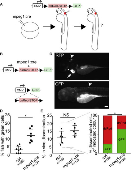
Macrophage Cytoplasmic Transfer to Melanoma Cells Correlates with Dissemination In Vivo (A and B) Strategy for Cre LoxP experiment, in which Cre is driven under a macrophage-specific promoter (mpeg1:Cre), and A375P human melanoma cells expressing CMV-LoxP-dsRed-STOP-LoxP-GFP are transplanted into transgenic larvae or control non-transgenics. Transfer of macrophage cytoplasm, including Cre recombinase, results in a switch in melanoma cell color from dsRed to GFP. (C) Lateral view of zebrafish larva with anterior to the left. Top panel shows RFP channel in which arrowhead points to tumor cell mass expressing dsRed. Red heart (arrow) indicates presence of mpeg1:Cre transgene. Bottom panel shows same larva in the GFP channel with single GFP+ disseminated tumor cell (arrowhead). Scale bar, 100 μm. (D) Quantification of the percent larvae with GFP+ tumor cells anywhere in the animal after transplantation into stable Tg(mpeg1:Cre)fh506 animals, compared with control non-transgenic animals. One-way ANOVA (p < 0.0001), followed by non-parametric unpaired t test, p = 0.003 (between ctrl and mpeg:cre). Error bars are mean ± SD. (E) Left side: quantification of percent larvae with any tumor cell dissemination (combining data for dsRed+ or GFP+) in stable Tg(mpeg1:Cre)fh506 animals, compared with control non-transgenic animals. Non-parametric unpaired t test, p = 0.10, not significant. Right side: ratio of disseminated tumor cells that are dsRed+ versus GFP+ in stable Tg(mpeg1:Cre)fh506 animals, compared with control non-transgenic animals. Non-parametric unpaired t test, p < 0.05 (between GFP+ cells in control and mpeg:cre). For (D) and (E), each data point is an independent experiment, n = number of larvae, and asterisks indicate p < 0.05. NS, not significant. Error bars are mean ± SD. See also Figure S5. |
Reprinted from Developmental Cell, 43, Roh-Johnson, M., Shah, A.N., Stonick, J.A., Poudel, K.R., Kargl, J., Yang, G.H., di Martino, J., Hernandez, R.E., Gast, C.E., Zarour, L.R., Antoku, S., Houghton, A.M., Bravo-Cordero, J.J., Wong, M.H., Condeelis, J., Moens, C.B., Macrophage-Dependent Cytoplasmic Transfer during Melanoma Invasion In Vivo, 549-562.e6, Copyright (2017) with permission from Elsevier. Full text @ Dev. Cell