
Fig. s3
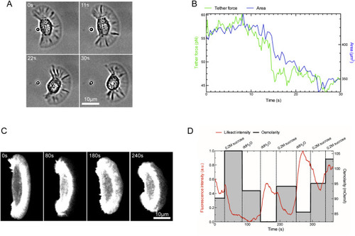
Membrane Tension Measurement by Tether Pulling and Membrane Tension Manipulation by Changing Osmotic Pressure, Related to Figure 2 (A) A migrating keratocyte was imaged with bright field microscopy and simultaneously a coated bead controlled by a laser tweezer was used to pull a membrane tether. (B) The measured tether force decrease correlated with rapid decreases in projected cell area. (C) A lifeact:GFP expressing keratocyte was imaged by confocal microscopy and subjected to rapid changes in osmotic pressure of the medium by alternating additions of 0.2M sucrose and pure water. (D) lifeact:GFP signal is negatively correlated with the osmolarity of the medium. When the pressure increases upon addition of 0.2M sucrose, lifeact:GFP signal drops, which is reversible upon restoring a lower osmotic pressure by addition of pure water. |
Reprinted from Cell, 171, Mueller, J., Szep, G., Nemethova, M., de Vries, I., Lieber, A.D., Winkler, C., Kruse, K., Small, J.V., Schmeiser, C., Keren, K., Hauschild, R., Sixt, M., Load Adaptation of Lamellipodial Actin Networks, 188-200.e16, Copyright (2017) with permission from Elsevier. Full text @ Cell