
Fig. 3
- ID
- ZDB-FIG-180302-15
- Publication
- Seritrakul et al., 2017 - Tet-mediated DNA hydroxymethylation regulates retinal neurogenesis by modulating cell-extrinsic signaling pathways
- Other Figures
- All Figure Page
- Back to All Figure Page
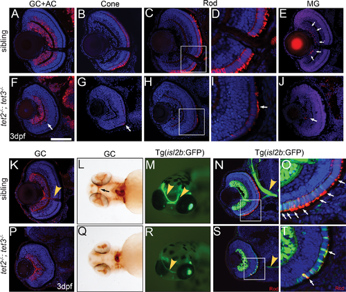
Fig 3. tet2-/-;tet3-/- retinal cells do not undergo terminal differentiation. (A,F) HuC/D labels RGCs and amacrine cells (ACs), which are reduced in number and located only in the central region of the INL in tet2-/-;tet3-/- retinae (arrow). (B,G) tet2-/-;tet3-/- mutant retinae almost entirely lack zpr-1+ red/green cones (arrow); (C,D,H,I) possess few zpr-3+ rods (arrow), and of those that are zpr-3+, outer segments are severely attenuated or almost absent (arrow). (E,J) tet2-/-;tet3-/- retinae also possess few zrf-1+ Müller glia (arrows in wild-type). In all cases, marker+ cells are located in the central/ventral part of the retina. (K,P) Zn8 detects neurolin, a protein enriched on RGCs and the optic nerve (arrowhead in K). (L,Q) Zn8 staining reveals the optic nerve in the choroid fissure and optic chiasm (arrow) of wild-type embryos but not in tet2-/-;tet3-/- mutants. (M-N) isl2b:GFP transgenics express GFP in RGCs and PRs, clearly labeling the optic nerve in whole-mount and section views (arrowhead). (R,S) The tet2-/-;tet3-/- optic nerve is very thin, often unilaterally formed, but, when present, correctly routed to the brain. (O,T) The isl2b:GFP signal overlaps zpr-3 (rod) marker in the cell body and outer segments in siblings (arrows). In tet2-/-;tet3-/-, few isl2b:GFP+ cells are zpr-3+ (arrows), further suggesting that specified cells are not terminally differentiated. Few outer segments have also formed in tet2-/-;tet3-/- mutants. DNA (blue), antibody stain (red). All images are 3dpf. n>5 for each marker. Dorsal is up and anterior to the left. Scale bar = 80μm in A-K, P. |