
Fig. 2
- ID
- ZDB-FIG-180302-14
- Publication
- Seritrakul et al., 2017 - Tet-mediated DNA hydroxymethylation regulates retinal neurogenesis by modulating cell-extrinsic signaling pathways
- Other Figures
- All Figure Page
- Back to All Figure Page
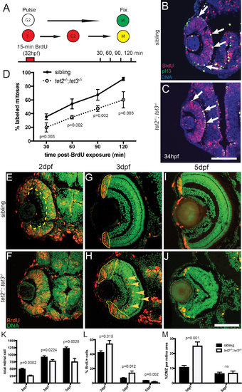
Fig 2. RPC cell cycle dynamics are disrupted in tet2-/-;tet3-/-. A) Percent labeled mitoses (PLM) assay was performed by treating embryos for 15 minutes in bromodeoxyuridine (BrdU) pulse at 32hpf, rinsed, fixed at 30, 60, 90, and 120 minutes post-treatment for immunostaining. (B,C) Cells in S-phase during BrdU pulse (BrdU+; red) and cells in G2/M-phase at fixation (pH3+; green) were counted. Cells that were proliferative during the BrdU pulse and then undergo mitosis are double positive (BrdU+,pH3+; yellow; arrows). (D) tet2-/-;tet3-/- retinae show significantly lower proportion of labeled mitotic events ([BrdU+pH3+]/pH3+) at all four time points. By the end of 120-minute window, nearly all ‘labeled’ RPCs completed S to M phase transition in wild-type siblings, compared to only 50% in tet2-/-;tet3-/- mutants (n = 5 embryos per condition per timepoint analyzed). (E-M) At later time points, BrdU incorporation assays over 2-hour time windows revealed that retinal progenitor cells remain proliferative longer and proliferate ectopically in tet2-/-;tet3-/- mutants. (E,F) At 2dpf, cells within the central retina of wild-type sibling embryos (dotted line) are no longer proliferative, correlating with cell cycle exit and differentiation. (G,H) At 3dpf, the only proliferative cells in the wild-type retina are located in the CMZ (outlined). In tet2-/-;tet3-/- mutants, this proliferative region is significantly expanded. Ectopically proliferating cells are also observed outside of the CMZ at significantly higher numbers than wild-type siblings (arrowheads). (I,J) At 5dpf, both sibling and tet2-/-;tet3-/- eyes possess proliferative CMZs. (K) Total retinal cell count per section is significantly lower in tet2-/-;tet3-/- mutants compared to siblings at all time points, correlating with microphthalmia. (L) tet2-/-;tet3-/- mutant retinae possess a significantly higher percentage of proliferative (BrdU+) cells at 2 and 3dpf, but lower at 5dpf. (M) Percentage of CMZ area per total retina is significantly higher in tet2-/-;tet3-/- mutant at 3dpf, but not significantly different at 5dpf. N = 3 embryos per condition per time point for E-M. Dorsal is up and anterior to the left in all images. All error bars = ± 1 S.D. All p-values calculated using two-tailed, unpaired t-test. Scale bar = 50μm in B-C; 80μm in E-J. |
| Fish: | |
|---|---|
| Observed In: | |
| Stage Range: | Prim-15 to Day 5 |