
Fig. S10
- ID
- ZDB-FIG-180206-8
- Publication
- Singh et al., 2017 - Different developmental histories of beta-cells generate functional and proliferative heterogeneity during islet growth
- Other Figures
- All Figure Page
- Back to All Figure Page
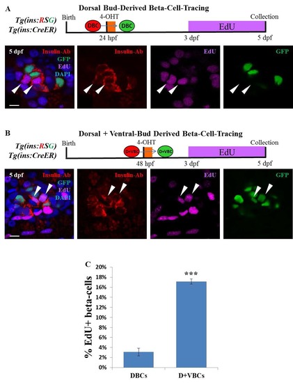
Analysis of beta-cell proliferation in DBCs and the combined D+VBCs population during an equivalent developmental window. (A) Top - schematic showing the rationale for studying the proliferation of DBCs. Tg(ins:mCherry-Stop-H2B-EGFP); Tg(ins:CreERT2) animals were incubated with 1 uM 4-OHT from 24 to 30 hpf in order to label specifically the DBCs with nuclear GFP fluorescence. The larvae were placed in 2.5 mM EdU from 3 to 5 dpf to label all proliferating beta-cells. Bottom - a single confocal section showing GFP-positive DBCs at 5 dpf. The DBCs (green) are EdU-negative. In contrast, the adjacent GFP-negative beta-cells are EdU-positive. Note that the red (mCherry)-fluorescence from Tg(ins:mCherry-Stop-H2B-EGFP)was lost due to the Click-iT reaction used to reveal EdU-incorporation. The GFP-signal was recovered using an anti-GFP antibody. All beta-cells were marked using an anti-insulin antibody. (B) Top - schematic showing the rationale for studying the proliferation of beta-cells in the combined D+VBCs population. Tg(ins:mCherry-Stop-H2B-EGFP); Tg(ins:CreERT2) embryos were incubated with 2 uM 4-OHT at 48 hpf in order to label the D+VBCs with nuclear GFP fluorescence. Larvae were placed in 2.5 mM EdU from 3 – 5 dpf to label all proliferating beta-cells. Bottom - a single confocal section showing GFP-positive beta-cells from the combined D+VBC population at 5 dpf. Arrowheads indicate EdU and GFP-double-positive beta-cells. Note that the red (mCherry)-fluorescence from Tg(ins:mCherry-Stop-H2B-EGFP) was lost due to the Click-it reaction used to reveal EdU-incorporation. The GFP-signal was recovered using an anti-GFP antibody. All beta-cells were marked using an anti-insulin antibody. (C) Quantification of the percentage of EdU and GFP-double-positive beta-cells from the experiments in A and B. The D+VBCs population contains a higher proportion of proliferating beta-cells as compared to the DBC population alone (unpaired t-test, *** p ≤ 0.001). n ≥ 5 islets at each stage with ≥10 GFP-positive cells per islet. Scale bars, 10 μm. |