
Fig. S12
- ID
- ZDB-FIG-180206-10
- Publication
- Singh et al., 2017 - Different developmental histories of beta-cells generate functional and proliferative heterogeneity during islet growth
- Other Figures
- All Figure Page
- Back to All Figure Page
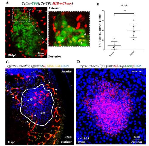
Lineage tracing of Notch-responsive cells. (A) Short-term lineage tracing of the Notch-responsive cells (NRCs) using histone-tagged mCherry. Maximum intensity projections of the primary islet from 18 dpf Tg(TP1:H2B-mCherry);Tg(ins:YFP) animals. Tg(TP1:H2B-mCherry)-expression labels the NRCs and Tg(ins:YFP) labels the beta-cells. The inset shows a higher magnification image of the anterior region of the islet. Due to the slow turnover of histone tagged proteins, Tg(Tp1:H2B-mCherry)+ progenitors retain the fused fluorescent protein upon differentiating into beta-cells, while at the same time they activate the insulin promoter. Thus, NRCs differentiating into beta-cells are co-labeled with YFP and H2B-mCherry (arrowheads in the inset). (B) Quantification of the short-term lineage tracing experiment. The plot shows the data points overlaid with a bar depicting Mean ± S.D. The anterior half of the islet harbors more YFP and H2B-mCherry-double positive cells as compared to the posterior half (t-test, ** p ≤ 0.01). (C) Uniform labeling of pancreatic ductal cells with Tg(Tp1:CreERT2). Maximum intensity projection of the pancreata from 21 dpf Tg(Tp1:CreERT2); Tg(-3.5ubb:loxP-EGFP-loxP-mCherry) animals incubated with 4-OHT at 4 dpf to induce recombination in the NRCs. Nkx6.1 immunofluorescence labels all ductal cells and DAPI labels cell nuclei. NRCs with successful recombination exhibit mCherry expression (red) and co-localize with the Nkx6.1+ cells (arrowheads) on all sides of the primary islet without any anterior/posterior bias. Note the presence of mCherry-positive and Nkx6.1-negative cells (arrow) within the anterior half of the primary islet (outlined by a white line), which likely represent NRCs that differentiated into the endocrine lineages. (D) Lack of leaky recombination in Tg(Tp1:CreERT2) animals at 30 dpf. Maximum intensity projection of the primary islets of Tg(Tp1:CreERT2); Tg(ins:mCherry-Stop-H2B-EGFP) animals incubated in vehicle at 4 dpf and stained with an anti-GFP antibody. Cells with leaky recombination would show green fluorescence. All samples (n = 15) lacked green fluorescence, suggesting absence of background recombination. Scale bars, 20 μm. |