
Fig. 2
- ID
- ZDB-FIG-180126-68
- Publication
- Pei et al., 2016 - Extracellular HSP60 triggers tissue regeneration and wound healing by regulating inflammation and cell proliferation
- Other Figures
- All Figure Page
- Back to All Figure Page
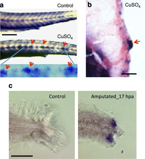
hspd1 expression is induced after injury. (a) hspd1 expression is induced in lateral line neuromasts by CuSO4-mediated hair cell ablation. Pictures are of embryos collected 5 h post-copper or control treatment, the time point with the largest expression differences. Arrows indicate the induced expression in lateral line neuromasts. The bottom panel is a higher magnification to more clearly show the neuromast-specific expression only seen in a CuSO4-treated embryo. (b) Histological sectioning shows the induced hspd1 expression in a cross-section of a neuromast localised in the trunk. (c) hspd1 expression is induced by caudal fin amputation. Pictures are of embryos collected at 17 h post amputation (or an undissected control), which was the peak of hspd1 expression. Bars = 200 μm in a and c, 20 μm in b. |
| Gene: | |
|---|---|
| Fish: | |
| Conditions: | |
| Anatomical Terms: | |
| Stage Range: | Protruding-mouth to Day 5 |