
Fig. 5
- ID
- ZDB-FIG-180122-6
- Publication
- Han et al., 2017 - Zygotic vinculin is not essential for embryonic development in zebrafish
- Other Figures
- All Figure Page
- Back to All Figure Page
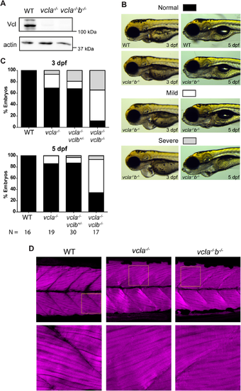
Cardiac and skeletal muscle phenotypes of vinculin-null mutants. (A) Western blot of lysates from the posterior half of WT, vcla-/- and vcla-/-vclb-/- embryos at 5 dpf, probed for vinculin and β-actin. (B) Some vinculin mutants show cardiac edema of which representative mild and severe cases are depicted. Wild type and vcla-/-vclb-/- mutants at 3 dpf (left) and 5 dpf (right). For corresponding images of the other genotypes described in (C), see supplemental S6 Fig. (C) Quantification of the presence of cardiac edemas in offspring from a vcla-/-vclb+/- incross. Classification as depicted in (B). Data was obtained from three independent experiments. WT embryos from an independent WT strain were analyzed as control from two independent experiments. Data is represented as mean ± s.e.m. A two-tailed paired student t-test was performed to compare the incidence of severe edemas between 3 dpf and 5 dpf within each genotype (seeS7 Fig). (D) Immunostaining of actin in skeletal muscle of 5 dpf embryos. Images at the bottom are zoomed in parts of the upper images as indicated by the yellow squares (see S8 Fig for additional images and quantifications). |
| Fish: | |
|---|---|
| Observed In: | |
| Stage Range: | Protruding-mouth to Day 5 |