
- Title
-
Zygotic vinculin is not essential for embryonic development in zebrafish
- Authors
- Han, M.K.L., van der Krogt, G.N.M., de Rooij, J.
- Source
- Full text @ PLoS One
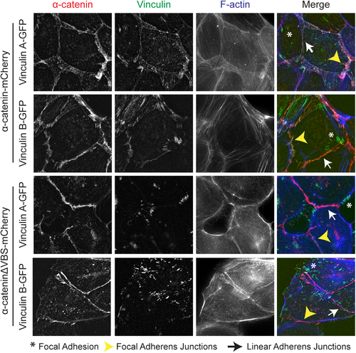
Zebrafish vinculin A and vinculin B localization. Fixed α-catenin-depleted MDCK epithelial cells expressing either α-catenin-mCherry (top two rows, depicted in red) or α-cateninΔVBS-mCherry, which lacks the vinculin binding domain (bottom two rows, depicted in red). In addition, cells express zebrafish vinculinA-GFP or vinculinB-GFP (both depicted in green) and were stained for F-actin (blue). Asterisks mark Focal Adhesions, White arrows mark Focal Adherens Junctions and Yellow Arrowheads mark Linear Adherens Junctions. |
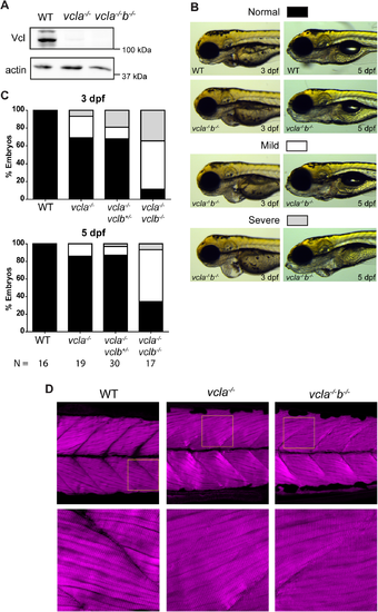
Cardiac and skeletal muscle phenotypes of vinculin-null mutants. (A) Western blot of lysates from the posterior half of WT, vcla-/- and vcla-/-vclb-/- embryos at 5 dpf, probed for vinculin and β-actin. (B) Some vinculin mutants show cardiac edema of which representative mild and severe cases are depicted. Wild type and vcla-/-vclb-/- mutants at 3 dpf (left) and 5 dpf (right). For corresponding images of the other genotypes described in (C), see supplemental S6 Fig. (C) Quantification of the presence of cardiac edemas in offspring from a vcla-/-vclb+/- incross. Classification as depicted in (B). Data was obtained from three independent experiments. WT embryos from an independent WT strain were analyzed as control from two independent experiments. Data is represented as mean ± s.e.m. A two-tailed paired student t-test was performed to compare the incidence of severe edemas between 3 dpf and 5 dpf within each genotype (seeS7 Fig). (D) Immunostaining of actin in skeletal muscle of 5 dpf embryos. Images at the bottom are zoomed in parts of the upper images as indicated by the yellow squares (see S8 Fig for additional images and quantifications). PHENOTYPE:
|
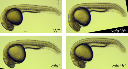
Offspring from incross of vcla-/-vclb+/-at 1 dpf. PHENOTYPE:
|
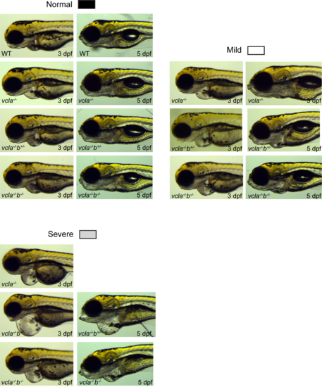
Pericardial edema in vinculin mutants at 3 dpf and 5 dpf. Pericardial edemas of embryos of the different vinculin genotypes were categorized by eye into normal, mild and severe on 3 and 5 dpf from three independent experiments. |

Skeletal muscle of vinculin mutants at 5 dpf. Representative images of skeletal muscle samples of wild-type control (A), vcla (B) and vcla/b double mutants (C) stained with phalloidin were analyzed at the intersomitic boundaries (bottom row). (D) quantification of the observed irregularities at the intersomitic boundaries. Data was obtained from two independent experiments. |

ZFIN is incorporating published figure images and captions as part of an ongoing project. Figures from some publications have not yet been curated, or are not available for display because of copyright restrictions. PHENOTYPE:
|