
FIGURE
Fig. 2
- ID
- ZDB-FIG-180122-5
- Publication
- Han et al., 2017 - Zygotic vinculin is not essential for embryonic development in zebrafish
- Other Figures
- All Figure Page
- Back to All Figure Page
Fig. 2
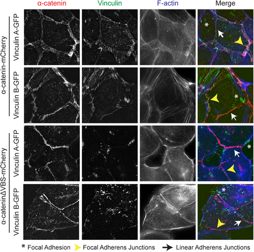
Zebrafish vinculin A and vinculin B localization. Fixed α-catenin-depleted MDCK epithelial cells expressing either α-catenin-mCherry (top two rows, depicted in red) or α-cateninΔVBS-mCherry, which lacks the vinculin binding domain (bottom two rows, depicted in red). In addition, cells express zebrafish vinculinA-GFP or vinculinB-GFP (both depicted in green) and were stained for F-actin (blue). Asterisks mark Focal Adhesions, White arrows mark Focal Adherens Junctions and Yellow Arrowheads mark Linear Adherens Junctions. |
Expression Data
Expression Detail
Antibody Labeling
Phenotype Data
Phenotype Detail
Acknowledgments
This image is the copyrighted work of the attributed author or publisher, and
ZFIN has permission only to display this image to its users.
Additional permissions should be obtained from the applicable author or publisher of the image.
Full text @ PLoS One