
FIGURE
Fig. 3
- ID
- ZDB-FIG-180111-2
- Publication
- Newman et al., 2017 - Mitochondrion to endoplasmic reticulum apposition length in zebrafish embryo spinal progenitors is unchanged in response to perturbations associated with Alzheimer's disease
- Other Figures
- All Figure Page
- Back to All Figure Page
Fig. 3
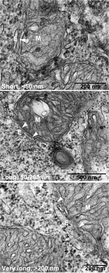
Electron microscopy of zebrafish neural cells. Cells were treated with morpholinos binding the start codon of psen1 and psen2 mRNA (MoPS1Tln and MoPS2Tln respectively). Arrowheads indicate the region of apposition between mitochondria (M) and endoplasmic reticulum (E) i.e. MAM. |
Expression Data
Expression Detail
Antibody Labeling
Phenotype Data
| Fish: | |
|---|---|
| Knockdown Reagents: | |
| Observed In: | |
| Stage: | Long-pec |
Phenotype Detail
Acknowledgments
This image is the copyrighted work of the attributed author or publisher, and
ZFIN has permission only to display this image to its users.
Additional permissions should be obtained from the applicable author or publisher of the image.
Full text @ PLoS One