
FIGURE
Fig. 2
- ID
- ZDB-FIG-180111-1
- Publication
- Newman et al., 2017 - Mitochondrion to endoplasmic reticulum apposition length in zebrafish embryo spinal progenitors is unchanged in response to perturbations associated with Alzheimer's disease
- Other Figures
- All Figure Page
- Back to All Figure Page
Fig. 2
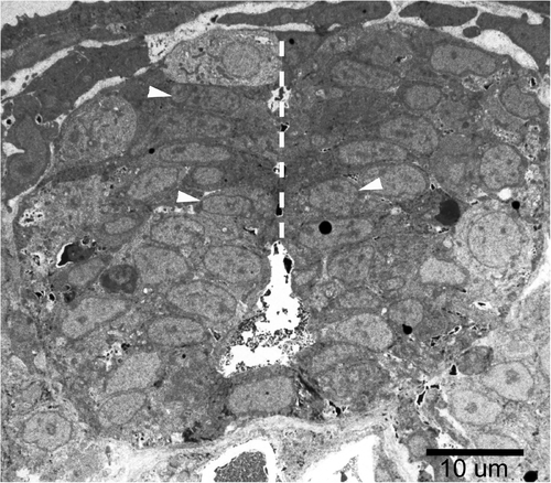
The midline of the spinal cord region. M-ER apposition lengths were measured in 3–6 cells at the midline area of the spinal cord at each embryo (such as those indicated by white arrowheads). Three embryos were examined for each treatment. The white, dashed line indicates the midline of the spinal cord. |
Expression Data
Expression Detail
Antibody Labeling
Phenotype Data
| Fish: | |
|---|---|
| Knockdown Reagents: | |
| Observed In: | |
| Stage: | Prim-5 |
Phenotype Detail
Acknowledgments
This image is the copyrighted work of the attributed author or publisher, and
ZFIN has permission only to display this image to its users.
Additional permissions should be obtained from the applicable author or publisher of the image.
Full text @ PLoS One