
Fig. S5
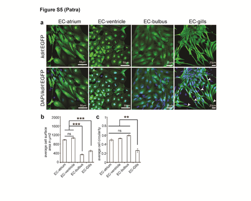
Morphological diversity of endothelial cells from different tissues. Cultured endothelial cells isolated from ventricle, atrium, bulbus arteriosus and gills. (a) Cells were stained for EGFP (endothelial cells, green), and DAPI (nuclei, blue) 2 days post seeding. White arrowheads point to elongated endothelial cells. (b, c) Quantitative analysis of the average surface area and average circularity of individual endothelial cells of different origins (n=3, mean±SEM). One way ANOVA followed by Bonferroni’s post-hoc test (GraphPad Prism) was performed to evaluate statistical significance of differences. P< 0.05 was considered statistically significant. *** corresponds to P<0.001 and ** corresponds to P<0.05. |