
Fig. S2
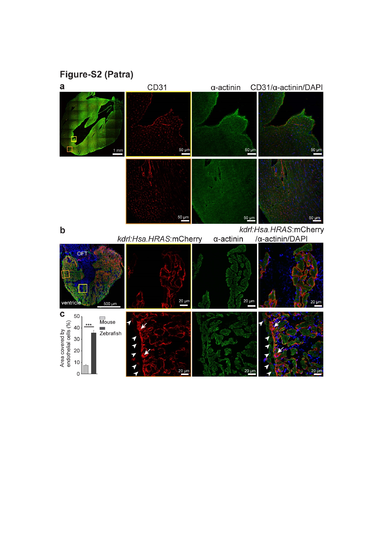
Endothelial cell density in 8 months old mouse and zebrafish ventricles. (a) Representative confocal images of sagittal cryosections through 8 months old mouse hearts stained for α-actinin (green; marking cardiomyocytes), CD31 (red; marking the cell membrane of endothelial cells) and DAPI (blue; staining the nuclei). (b) Representative confocal images of sagittal cryosections through 8 mpf Tg(kdrl:Hsa.HRAS-mCherry) zebrafish hearts stained for α-actinin (green; marking cardiomyocytes), mCherry (red; marking endothelial cell membrane) and DAPI (blue; staining the nuclei). White arrowheads point to bigger coronary vessels in the outer compact layer; white arrows point to dense capillaries in the inner compact layer. (c) Quantification of the area covered by endothelial cells (as percentage of total cardiac tissue area). 10 sections from each heart from three zebrafish and three mice were analyzed (mean±SEM). One way ANOVA followed by Bonferroni’s post-hoc test (GraphPad Prism) was performed to evaluate statistical significance of differences. p < 0.05 was considered statistically significant. *** corresponds to P<0.001. |