
Fig. 1
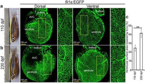
Zebrafish coronary vessels continue to grow after 3 months post fertilization. (a,b) Representative brightfield images of adult zebrafish and maximum intensity projections of confocal images of freshly isolated hearts from 110 (a) and 230 (b) dpf Tg(fli1a:EGFP) animals. Green cells have pan-endothelial identity. (c) Quantification of cardiac ventricular surface area covered by endothelial cells (n = 3 of each stage, mean ± SEM). One way ANOVA followed by Bonferroni’s post-hoc test (GraphPad Prism) was performed to evaluate statistical significance of differences. P < 0.05 was considered statistically significant. ** corresponds to P < 0.05. AVC- atrioventricular canal, dpf- days post fertilization. |