
Fig. 8
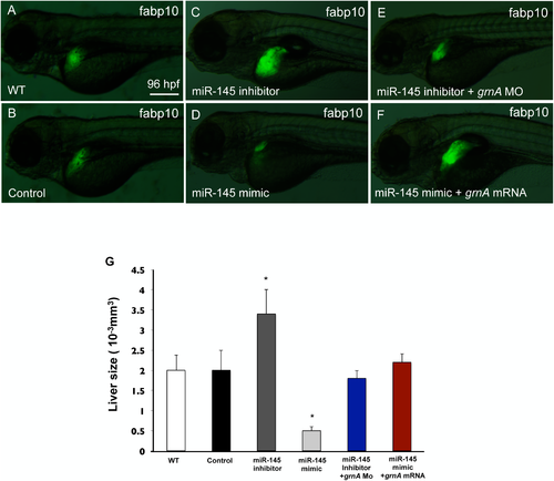
GrnA rescues the hepatic outgrowth defect caused by miR-145 manipulation. Liver morphology was determined by EGFP expression at 4 dpf in Tg (fabp10:EGFP) embryos (A) or in embryos injected with control mimic (B), miR-145 inhibitor (C), miR-145 mimic (D), miR-145 inhibitor with grnA MO (0.25 ng/embryo) (E) and miR-145 mimic with grnA mRNA (0.4 ng/embryo) (F). Thirty embryos per experimental group were used and three independent replicates were performed. A 3D image of the liver was observed using Leica SP5 confocal microscope and Imaris software. The liver size was examined by measuring the volume of EGFP expression (G). Ten embryos per experimental group were used and three independent replicates were performed. Scale bars, 100 μm; EGFP, enhanced green fluorescent protein; *P < 0.05, t-test. |