
Fig. S7
- ID
- ZDB-FIG-171227-16
- Publication
- Shao et al., 2017 - Vegetally localised Vrtn functions as a novel repressor to modulate bmp2b transcription during dorsoventral patterning in zebrafish.
- Other Figures
- All Figure Page
- Back to All Figure Page
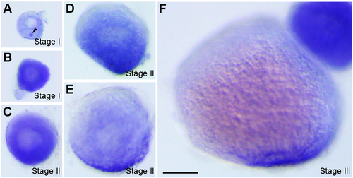
Localisation of vrtn transcripts during oogenesis. (A) Stage I oocyte (80 μm in diameter), with vrtn transcripts detected in the ooplasm, and in the Balbiani body (arrowhead). (B) Stage I oocyte (100 μm in diameter), with strong uniform localisation in the ooplasm. (C) Stage II oocyte (150 μm in diameter) shows a slight vegetal enrichment. (D) Stage II oocyte (200 μm in diameter), with evident vegetal localisation. (E) Stage II oocyte (230 μm in diameter) shows vrtn localisation mainly in the vegetal region. (F) Stage III oocyte with vrtn transcripts restricted exclusively in the vegetal pole cortex. Oocytes were staged according to their diameters: stage I, 70-140 μm; stage II, 140-340 μm; stage III, 340-690 μm. Scale bar: 100 μm. |
| Gene: | |
|---|---|
| Fish: | |
| Anatomical Terms: | |
| Stage: | Adult |