
Fig. 1
- ID
- ZDB-FIG-171101-2
- Publication
- Chen et al., 2015 - Nmnat1-Rbp7 Is a Conserved Fusion-Protein That Combines NAD+ Catalysis of Nmnat1 with Subcellular Localization of Rbp7
- Other Figures
- All Figure Page
- Back to All Figure Page
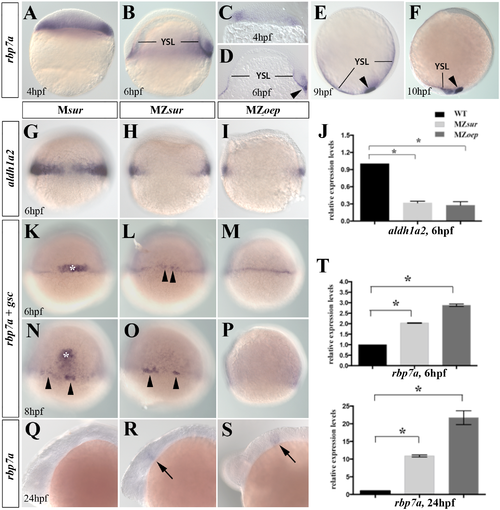
Complementary regulation of rbp7a and aldh1a2 by Nodal signaling. [A-F] Lateral views of whole mount in situ stains of rbp7a in wild type embryos at 4hpf [A, C] 6hpf [B,D], 9hpf [E] and 10hpf [F]; [C-D] vibratome sections of 4hpf and 6hpf embryos. rbp7a signals are found in marginal cell [A-D], YSL, and in forerunner cells (arrowhead in [D-F]). [G-J] Marginal expression of aldh1a2 is strongly reduced in 6hpf MZsur [H] and MZoep [I] embryos; [J] RT-qPCR verified reduced expression levels of aldh1a2 in MZsur, MZoep as compared to wild type embryos. [K-P] Whole mount in situ hybridization for rbp7a (arrow heads) and goosecoid (gsc) (white asterisks) performed at 6 and 8 hpf in Msur+/- as control [K, N], MZsur [L, O] and MZoep [M, P] embryos. Note that expression levels of rbp7a around the YSL were similar among the gsc positive embryos and the gsc negative MZsur [B, E] and MZoep [C, F] mutants. [Q-S] rbp7a expression at 24hpf. Arrows mark rbp7a signals in the posterior midbrain that were present in MZsur [R] and MZoep embryos [S] but not in control embryo [Q]. [T] RT-qPCR results for rbp7a in of 6hpf and 24hpf embryos. Graphed is the mean and SEM from triplicate experiments. Error bars indicated the SEM. Unpaired T-test was used to test the significance (*P<0.05). |
| Genes: | |
|---|---|
| Fish: | |
| Anatomical Terms: | |
| Stage Range: | Sphere to Prim-5 |
| Fish: | |
|---|---|
| Observed In: | |
| Stage Range: | Shield to Prim-5 |