
FIGURE
Fig. 7
- ID
- ZDB-FIG-171030-38
- Publication
- Lee et al., 2015 - Association of Early Atherosclerosis with Vascular Wall Shear Stress in Hypercholesterolemic Zebrafish
- Other Figures
- All Figure Page
- Back to All Figure Page
Fig. 7
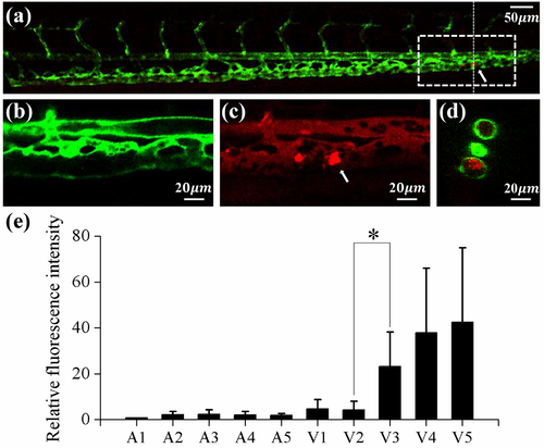
Lipid accumulation along the main blood vessel of 15 dpf zebrafish. (a) Typical confocal microscopy image of the caudal vasculature in a 15 dpf zebrafish. Bright fluorescent displays lipid deposit in the blood vessel. White arrows denote the location of lipid accumulation. ECs and fluorescent lipid image in the square dotted region were magnified in (b) and (c), respectively. (a)~(c) 2D sectional image, lateral view. (d) Cross sectional image at the vertical dotted line in (a). (e) Variation of relative fluorescence intensity of lipid in10 vessels. (n = 11) *P < 0.001. |
Expression Data
Expression Detail
Antibody Labeling
Phenotype Data
| Fish: | |
|---|---|
| Condition: | |
| Observed In: | |
| Stage: | Days 14-20 |
Phenotype Detail
Acknowledgments
This image is the copyrighted work of the attributed author or publisher, and
ZFIN has permission only to display this image to its users.
Additional permissions should be obtained from the applicable author or publisher of the image.
Full text @ PLoS One