
FIGURE
Fig. 3
- ID
- ZDB-FIG-170823-3
- Publication
- Walderich et al., 2016 - Homotypic cell competition regulates proliferation and tiling of zebrafish pigment cells during colour pattern formation
- Other Figures
- All Figure Page
- Back to All Figure Page
Fig. 3
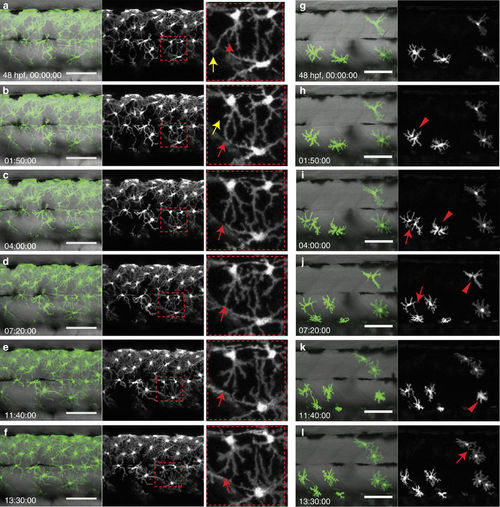
Interaction of xanthophores in larval stage. (a–f) Live-imaging of wild-type xanthophores—green; Tg(pax7:GFP), shows that cell numbers remain constant during larval development. (Same images in grey scale). Magnification of red dotted boxes: dynamic cell–cell contacts occur by extension (red arrows) and retraction (yellow arrows) of filopodia. (g–l) Live-imaging of xanthophores—green; Tg(pax7:GFP) in pfeffer chimera. Transplanted xanthophores divide, for better visibility same image in grey scale (red arrowheads) and extent filopodia to each other (red arrow). Scale bars, a–l: 100 μm. |
Expression Data
Expression Detail
Antibody Labeling
Phenotype Data
Phenotype Detail
Acknowledgments
This image is the copyrighted work of the attributed author or publisher, and
ZFIN has permission only to display this image to its users.
Additional permissions should be obtained from the applicable author or publisher of the image.
Full text @ Nat. Commun.