
Fig. 1
- ID
- ZDB-FIG-170823-1
- Publication
- Walderich et al., 2016 - Homotypic cell competition regulates proliferation and tiling of zebrafish pigment cells during colour pattern formation
- Other Figures
- All Figure Page
- Back to All Figure Page
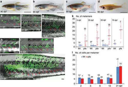
Development of xanthophore clusters in wild type and pfeffer. (a–d) Stripe pattern in 3-month-old (a) wild-type zebrafish, (b) pfeffer mutant, (c) chimera obtained by blastomere transplantation of wild type (Tg(pax7:GFP)) into pfeffer; the area with donor xanthophores is demarcated by a red dashed line and (d) control (albino). In (a) the dark stripes nomenclature is depicted along the dorsoventral axis. Developmental profile of Tg(pax7:GFP)-labelled wild-type xanthophores (green) in a (e–g) control and (h–j) pfeffer chimera. Dashed white lines—vertical and horizontal myosepta. Scale bars, a–d=1 cm; e–j=100 μm. (k) Quantification of the number of metameres spanned by xanthophore clusters at larval stage (6 dpf) (wild type, n=19 clusters, 12 fishes; pfeffer, n=30 clusters, 18 fishes; P≤0.0001); 26 dpf (wild type, n=37 clusters, 10 fishes; pfeffer, n=38 clusters, 17 fish; P≤0.0001); 33 dpf (wild type, n=47 clusters, 10 fishes; pfeffer, n=35 clusters, 17 fishes; P<0.0001); 70 dpf (wild type, n=7 clusters, 4 fishes; pfeffer, n=7 clusters, 7 fishes; P=0.0077). The horizontal lines in dark blue (wild type) and red (pfeffer) indicate the mean value and the error bars represent standard deviation. We show that the differences between albino and pfeffer are significant at all time points investigated by using Student’s t-test (Welsh corrected). (l) Number of xanthophores per metamere in wild type (blue) and pfeffer transplants (red). Numbers of metameres analysed are depicted in the graph, error bars represent standard deviation. No significant differences between wild-type fish (blue) and pfeffer transplants (red) according to Student;s t-test (Welsh corrected) were found for 2 dpf: P=0.1795 and 21 dpf: P=0.6123. Overall 3 dpf (P=<0.0001), 6 dpf (P=<0,0001) and 13 dpf (P=0.0042) were shown to be significantly different. |