
FIGURE
Fig. S5
- ID
- ZDB-FIG-170706-44
- Publication
- Tekeli et al., 2017 - Fate predetermination of cardiac myocytes during zebrafish heart regeneration
- Other Figures
- All Figure Page
- Back to All Figure Page
Fig. S5
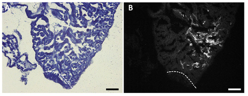
Labeled CMs close to the injury site. Sections processed for Sudan black staining showing the closest labeled cardiomyocytes to the injury site (a,b) in which GFP-positive cardiomyocytes did not contribute to the regenerated tissue. (a) Bright field image and (b) GFP positive clone. This heart corresponds to the regenerated heart in the panel q and q' from the Supplementary figure S4. The amputation plane is indicated by dashed lines. Scale bars: 250μm |
Expression Data
Expression Detail
Antibody Labeling
Phenotype Data
Phenotype Detail
Acknowledgments
This image is the copyrighted work of the attributed author or publisher, and
ZFIN has permission only to display this image to its users.
Additional permissions should be obtained from the applicable author or publisher of the image.
Full text @ Open Biol.