
Fig. 4
- ID
- ZDB-FIG-170706-40
- Publication
- Tekeli et al., 2017 - Fate predetermination of cardiac myocytes during zebrafish heart regeneration
- Other Figures
- All Figure Page
- Back to All Figure Page
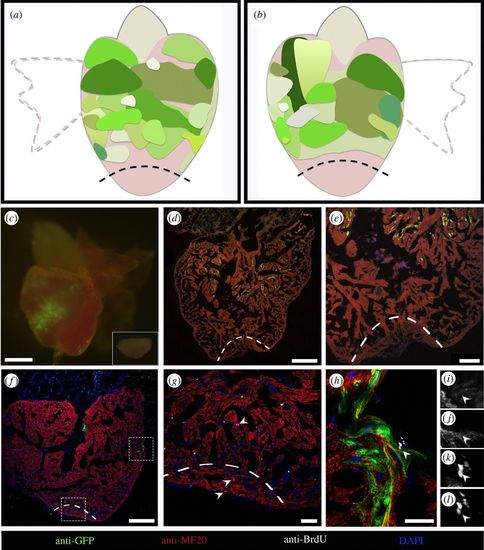
CMs distant from the amputation site do not contribute to the regenerated tissue. (a,b) Diagram summarizing the size and location of GFP-positive areas from different hearts which did not contribute to regeneration (n = 15). (a) The dorsal view of a zebrafish heart and (b) the ventral view. (c) Epifluorescence image of a representative heart where the amputation plane did not pass through the GFP-positive area. The excised portion of tissue is shown in inset. (d,e) Collected hearts were sectioned, and processed for immunofluorescence with antibodies against MHC and GFP. GFP-labelled CMs are absent in the regenerated area. (f–h) Sections processed for immunofluorescence with antibodies against BrdU, GFP and MHC showing the proliferating CMs (arrowheads) in the injury site (g) and in the GFP-positive area (h). Single image channels of the BrdU-positive GFP-positive CM (arrowhead) shown in (h), where (i) is GFP, (j) is MHC, (k) is DAPI and (l) is BrdU. Magnification images of lower and upper dashed rectangles in (f) are shown in (g) and (h), respectively. The amputation plane is indicated by dashed lines. Nuclei were counterstained with DAPI. Scale bars: (c) 500 µm, (d,f) 250 µm, (e) 100 µm, (g) 40 µm and (h)15 µm. |