
Fig. 6
- ID
- ZDB-FIG-170609-5
- Publication
- Ding et al., 2016 - MicroRNA Profiling during Craniofacial Development: Potential Roles for Mir23b and Mir133b
- Other Figures
- All Figure Page
- Back to All Figure Page
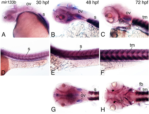
Figure 6. Expression of mir133b in zebrafish embryos. Whole-mount in situ hybridization analysis with a digoxigenin-labeled probe against mir133b at 30–72 hpf. (A–C) Expression is observed in the developing pharyngeal region, including at 48 hpf in the facial muscle (fm) and otic vessicle (B,G). (D–F) Strong expression of miR133b is observed in the somites (s; D) at 30 hpf, somites (E), and pectoral fin (pf) at 48 hpf (H) and facial muscle (fm; C), cardiac muscle (cm; C), and trunk muscle (tm; F) at 72 hpf, including the anterior mandibularis (arrows) and stemohyoideus (arrowheads) facial muscles muscles. Anterior is to the left. e, eye; tm, trunk muscle; n, notochord; ch, ceratohyal. |
| Gene: | |
|---|---|
| Fish: | |
| Anatomical Terms: | |
| Stage Range: | Prim-15 to Protruding-mouth |