
FIGURE
Fig. S5
- ID
- ZDB-FIG-170607-26
- Publication
- Benedetti et al., 2016 - INaP selective inhibition reverts precocious inter- and motorneurons hyperexcitability in the Sod1-G93R zebrafish ALS model
- Other Figures
- All Figure Page
- Back to All Figure Page
Fig. S5
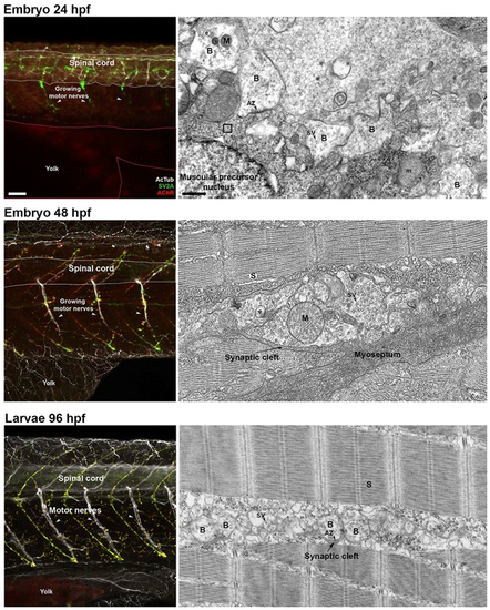
Morphological and ultrastructural changes in developing zebrafish locomotor network. Control (Ctrl) embryos 24 hpf (upper panels) show short motor axons protruding from the spinal cord entirely filled with synaptic vesicles (stained with anti-SV2A antibodies - green). This outgrowth has a rostral to caudal developmental pattern, and only the more rostral motor nerves show branches. Acetylated tubulin (grey) stains a few spinal interneuronal axonal projections. At this developmental stage, no clusters of acetylcholine receptors (AChRs - red) are visible on muscle fibre precursors. Scale bar: 20 µm. The ultrastructural analysis confirmed that the axonal projections are filled with synaptic vesicles (sv), with immature boutons (B) facing muscle fibres precursors with glycogen-filled cytoplasm (square box) and a poorly organized contractile apparatus. Scale bar: 500 nm. By 48 hpf (middle panels), the motor nerves present a well-organized microtubule network along their entire length, deeply penetrating the trunk along myosepta, and begin innervating the branched muscle fibre precursors. The axons are no longer entirely filled with synaptic vesicles, which are well organized in small clusters at the tips of the axonal branches, and the muscle fibres begin to show visible clusters of AChRs (red). Electron micrographs show pre-synaptic terminals filled with vesicles (sv), mainly located at the periphery of myotomes, and muscle fibres with a well-organized contractile apparatus (S). By 96 hpf (lower panels), the larvae show well-developed, heavily branched motor nerves innervating muscle fibres. Synaptic vesicles are distributed in small clusters at the tips of the axonal terminals, which now face the AChR clusters on muscle fibers (their superimposition generates a yellow signal in the merged image). Ultrastructural analysis reveals small pre-synaptic boutons (B) deeply penetrating into the myotome and innervating well-developed muscle fibres. The confocal images are maximum projections of z-stacks covering half of the trunk. Symbols: AZ: active zone; M: pre-synaptic mitochondria; m: muscle mitochondria; e: endosome. |
Expression Data
Expression Detail
Antibody Labeling
Phenotype Data
Phenotype Detail
Acknowledgments
This image is the copyrighted work of the attributed author or publisher, and
ZFIN has permission only to display this image to its users.
Additional permissions should be obtained from the applicable author or publisher of the image.
Full text @ Sci. Rep.