
Fig. 1
- ID
- ZDB-FIG-170607-17
- Publication
- Benedetti et al., 2016 - INaP selective inhibition reverts precocious inter- and motorneurons hyperexcitability in the Sod1-G93R zebrafish ALS model
- Other Figures
- All Figure Page
- Back to All Figure Page
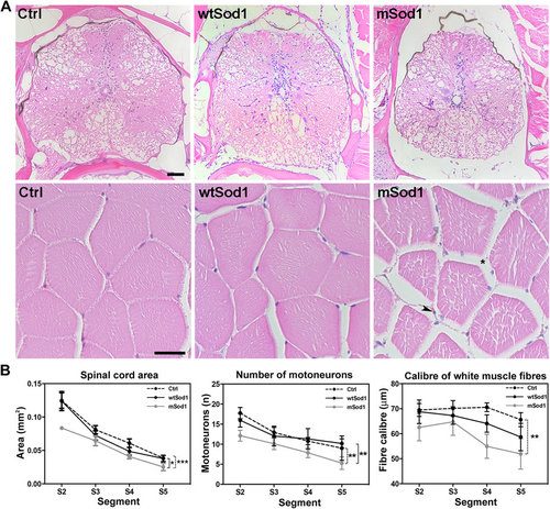
Spinal cord and lateral muscle histological analyses. Each adult fish was transversely cut into five segments (S1–S5) using the fins as anatomical references (see Supp. Figure S2). (A) Hematoxylin & eosin-stained histological sections of the S2 segment of the spinal cord (upper panels; scale bar: 50 μm) and white muscle fibres (lower panels; scale bar: 25 μm) in control (Ctrl), wtSod1 and mSod1 zebrafish. Muscular atrophy and edema (*) with infiltrating cells (arrowhead) are visible in mSod1 lateral muscle. (B) The plots show the significant reduction in spinal cord area and the number of motor neurons throughout the spinal cord, and a significant reduction in the calibre of white muscle fibres along the mSod1 fish trunk. Each point in the plots shows the mean value ± SEM of the indicated parameter in each segment of seven animals for each genotype. The measures were statistically analyzed using two-way ANOVA, and corrected by means of Sidak’s multiple comparison test (*P < 0.05; **P < 0.01; ***P < 0.001). |
| Fish: | |
|---|---|
| Observed In: | |
| Stage: | Adult |