
FIGURE
Fig. 3
- ID
- ZDB-FIG-170512-33
- Publication
- Kainrath et al., 2017 - Green-Light-Induced Inactivation of Receptor Signaling Using Cobalamin-Binding Domains
- Other Figures
- All Figure Page
- Back to All Figure Page
Fig. 3
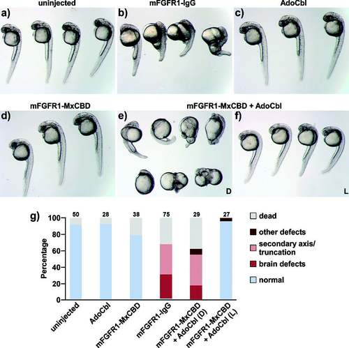
Embryos a) not injected (n.i.) or injected at the one-cell stage with b) constitutively active mFGFR1-IgG (13 pg plasmid), c) AdoCbl (50 fmol), d) mFGFR1-MxCBD (13 pg plasmid), e) mFGFR1-MxCBD (13 pg plasmid) and AdoCbl (25 fmol) raised in the dark (D), and f) mFGFR1-MxCBD (13 pg plasmid) and AdoCbl (25 fmol) raised under green light irradiation (L; 545±5 nm, I=180 μW cm−2 from 1 to 24 hpf). The images were recorded after 24 (a, c, d) and 30 hpf (b, e, f). g) Quantification of phenotypes (numbers denote the number of embryos). |
Expression Data
Expression Detail
Antibody Labeling
Phenotype Data
Phenotype Detail
Acknowledgments
This image is the copyrighted work of the attributed author or publisher, and
ZFIN has permission only to display this image to its users.
Additional permissions should be obtained from the applicable author or publisher of the image.
Full text @ Angew. Chem. Int. Ed. Engl.