
- Title
-
Green-Light-Induced Inactivation of Receptor Signaling Using Cobalamin-Binding Domains
- Authors
- Kainrath, S., Stadler, M., Reichhart, E., Distel, M., Janovjak, H.
- Source
- Full text @ Angew. Chem. Int. Ed. Engl.
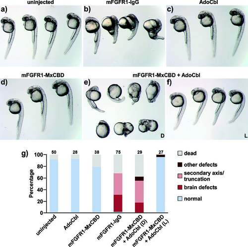
Embryos a) not injected (n.i.) or injected at the one-cell stage with b) constitutively active mFGFR1-IgG (13 pg plasmid), c) AdoCbl (50 fmol), d) mFGFR1-MxCBD (13 pg plasmid), e) mFGFR1-MxCBD (13 pg plasmid) and AdoCbl (25 fmol) raised in the dark (D), and f) mFGFR1-MxCBD (13 pg plasmid) and AdoCbl (25 fmol) raised under green light irradiation (L; 545±5 nm, I=180 μW cm−2 from 1 to 24 hpf). The images were recorded after 24 (a, c, d) and 30 hpf (b, e, f). g) Quantification of phenotypes (numbers denote the number of embryos). |
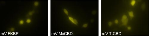
Representative fluorescence microscopy images for mV-FKPB, mV-MxCBD, and mV-TtCBD. |