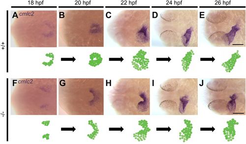
Homozygous pdgfra mutants demonstrate abnormal cardiac fusion. Representative photomicrographs of whole-mount in situ hybridization using the cardiac myosin light chain 2 (cmlc2) riboprobe to label myocardial cells. In +/+ embryos, the posterior regions of the bilateral cardiac fields interact during cardiac fusion at 18 hpf (A), followed by interaction between the anterior regions at 20 hpf (B). At 22 hpf, the cardiac tube forms and symmetry is broken by leftward displacement (jogging) (C). At 24 (D) and 26 (E) hpf, the cardiac tube elongates. In −/− mutants, cardiac fusion is delayed at 18 hpf (F). Interaction between the posterior regions occurs at 20 hpf (G). At 22 hpf, the anterior regions of the bilateral cardiac fields fail to fuse (H). By 24 (I) and 26 (J) hpf, the anterior portions of the bilateral cardiac fields begin to come into contact. Dorsal views are shown, with the fish cranium on the left. An illustration of the developing heart is shown at the bottom of each figure. Scale bar: 100 μm.
|
