
Fig. 3
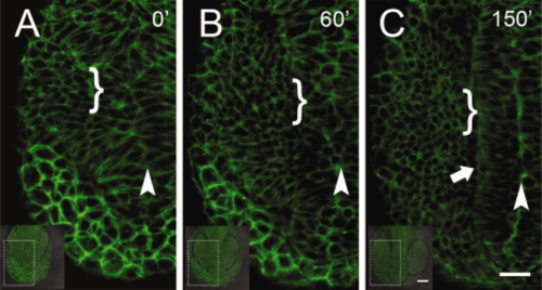
Analysis of early cell movements reveals a continuous field of B-actin:Gap43-GFP expressing cells. A–C: Initially, at 4 somite stage (ss), it is possible to identify the region of the forming neural tube lumen (A, arrowhead) but not the lateral neural tube which becomes well defined by 6–7 ss (C, arrowhead). The region of the pollster (anterior prechordal plate) initially (A,B) showed the most intense Bactin:GFP expression. The OP precursors lie very dorsal, and by 9–10 ss (C) become very round in shape (brackets). Dorsal view, anterior toward bottom of page. GFP, green fluorescent protein. Scale bars = 50 µm in insets; 25 µm in A–C (see Supp. Movie S1). |