Fig. 2
|
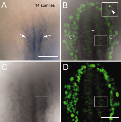
Expression of emx1 colocalizes with Dlx3b in the developing telencephalon. A: Transmitted light image of whole-mount embryo processed for emx1 expression marking the telencephalon (arrowheads). B–D: Different preparation of 14 ss whole-mount embryo with Dlx3b immunodetection (B, D green) after in situ hybridization for emx1 (B,C, dark gray). Merged image of 3 µm focal plane (B) colocalizes Dlx3b signal (green) with transmitted light image of emx1 in situ (gray). Inset box in B shows higher magification of cells expressing Dlx3b (asterisk) and emx1 (arrowhead). All embryos are dorsal views with anterior toward top of page. OP, olfactory placodes; T, telencephalon. Scale bars = 100 μm in A; 50 μm in B–D. |

