
FIGURE
Fig. 1
- ID
- ZDB-FIG-170308-26
- Publication
- Huckenpahler et al., 2016 - Imaging the adult zebrafish cone mosaic using optical coherence tomography
- Other Figures
- All Figure Page
- Back to All Figure Page
Fig. 1
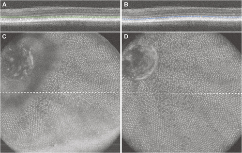
Visualization of the zebrafish cone mosaic using en face volume projection images. (A, C) The straight slab method in the native Bioptigen analysis software results in an en face volume projection with different retinal layers included at different locations within the volume, thus the cone mosaic is only visible in some parts of the en face image. (B, D) Using a custom contour that follows a single outer hyper-reflective band results in an en face projection of that layer, in this case the UV cone mosaic. |
Expression Data
Expression Detail
Antibody Labeling
Phenotype Data
Phenotype Detail
Acknowledgments
This image is the copyrighted work of the attributed author or publisher, and
ZFIN has permission only to display this image to its users.
Additional permissions should be obtained from the applicable author or publisher of the image.
Full text @ Vis. Neurosci.