
Fig. 1 S2
- ID
- ZDB-FIG-170223-28
- Publication
- Dyballa et al., 2017 - Distribution of neurosensory progenitor pools during inner ear morphogenesis unveiled by cell lineage reconstruction
- Other Figures
- All Figure Page
- Back to All Figure Page
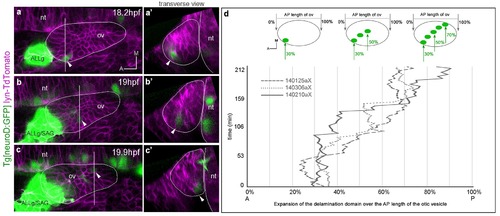
Posterior expansion of the otic neuroblast delamination domain. Tg[neuroD:GFP] embryos were injected with lyn-TdTomato mRNA at 1cell-stage and imaged from 14.5 hpf onwards. Embryos express GFP (green) in neuronal progenitors and differentiating neuroblasts, and TdTomato in all cell membranes (magenta). In the case of the inner ear, GFP is expressed in epithelial neuroblasts just prior to delamination and in the SAG neuroblasts. (a–c) Still image views of the ventral otic vesicle at the indicated time points showing the quick expansion of the delamination domain in the otic epithelium within 2 hr from anterolateral to posteromedial regions; note that at this stage the rudiment of the adjacent ALLg is already visible. (a’–c’) Transverse views are digital reconstructions along the lines indicated in (a–c) and illustrate that the onset of neuroblasts’ delamination progresses from lateral to medial domains (see arrowheads). ALLg, anterior lateral line ganglion; SAG, statoacoustic ganglion; nt, neural tube. The otic vesicle contour is depicted in white. ID Dataset: 140210aX. (d) Plot depicting the posterior expansion of the neuroblast delamination domain as assessed by neuroD-GFP expression. The position of posterior-most neuroD-GFP expressing cells in the otic epithelium, and the anterior and posterior edge of the otic vesicle (dorsal view) were assessed over time (see scheme). The plot displays the position of posterior-most GFP epithelial cells as a percent of the AP otic vesicle length (ID datasets: 140306aX, 140125aX, 140210aX). |