
Fig. S3
- ID
- ZDB-FIG-170209-21
- Publication
- Sotolongo-Lopez et al., 2016 - Genetic Dissection of Dual Roles for the Transcription Factor six7 in Photoreceptor Development and Patterning in Zebrafish
- Other Figures
- All Figure Page
- Back to All Figure Page
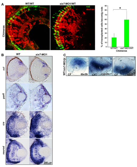
six7 functions in photoreceptor progenitor cells. (A) Histological sections of chimera retinas labeled for rods (4C12, green). six7-MO donor cells (red) preferentially generate rods compare to WT donor cells. Note the gap in rod labeling in WT/WT controls. Graph represents the percentage of donor cells that differentiate as rods in the central retina of WT/WT (n = 5) and six7-MO1/WTchimeras (n = 6). *p<0.05, student t test. (B) rx1-, pax6a-, crx- and neurod-in situ hybridization (blue) in a retinal cryosection from 48-hpf WT and six7-MO1 embryos. No labeling of retinal progenitors cell markers (rx1 and pax6a) were observed in the ONL of six7-morphant retinas. (C) Whole-mount in situ hybridization for the dorsal (tbx2b), midline (cyp26c1) and ventral (vax2) retinal marker. Labeling was indistinguishable different between WT and mutant embryos. |