
FIGURE
Fig. 2
- ID
- ZDB-FIG-170130-9
- Publication
- Du et al., 2016 - Spatial and Temporal Distribution of Dopaminergic Neurons during Development in Zebrafish
- Other Figures
- All Figure Page
- Back to All Figure Page
Fig. 2
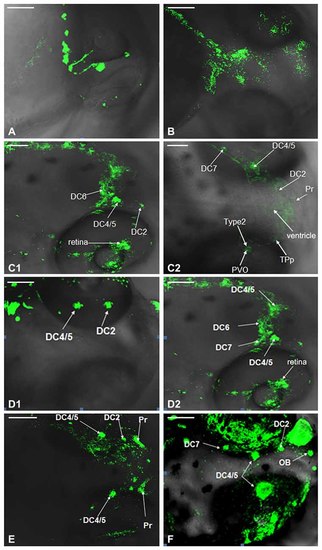
Development of dopaminergic neurons in wild type (WT) zebrafish. Development of dopaminergic neruons in WT zebrafish was determined by tyrosine hydroxylase (TH) whole mount immunofluorescent at 24 hpf (A), 48 hpf (B), 3 dpf (C1,C2), 4 dpf (D1,D2), 5 dpf (E) and 6 dpf (F), respectively. In order to show that it was not an individual case, we provided (C1,C2), and there was no difference between them. It was the same with (D1,D2). DC, diencephalons; Pr, pretectum; TPp, periventricular nucleus of posterior tubercle; PVO, paraventricular organ; OB, olfactory bulb. Scale = 100 μm. |
Expression Data
Expression Detail
Antibody Labeling
Phenotype Data
Phenotype Detail
Acknowledgments
This image is the copyrighted work of the attributed author or publisher, and
ZFIN has permission only to display this image to its users.
Additional permissions should be obtained from the applicable author or publisher of the image.
Full text @ Front. Neuroanat.