
- Title
-
Spatial and Temporal Distribution of Dopaminergic Neurons during Development in Zebrafish
- Authors
- Du, Y., Guo, Q., Shan, M., Wu, Y., Huang, S., Zhao, H., Hong, H., Yang, M., Yang, X., Ren, L., Peng, J., Sun, J., Zhou, H., Li, S., Su, B.
- Source
- Full text @ Front. Neuroanat.

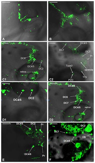
Development of dopaminergic neurons in Vmat2:GFP zebrafish. The development of dopaminergic neurons in ventral telencephalic area was observed repeatedly at 24 h post fertilization (hpf; A1–A3), 30 hpf (B1–B3), 48 hpf (C1–C3), 54 hpf (D1–D3), 3 days post fertilization (dpf; E1–E3), 4 dpf (F1–F3), 5 dpf (G1–G3). Similar observations are demonstrated in (A1–G3). DC, diencephalons; TPp, periventricular nucleus of posterior tubercle; MFB, midbrain/forebrain boundary; Vd/Vv, dorsal/ventral nucleus of the telencephalic area; LC, locus coeruleus; Ra, raphe nuclei; Pr, pretectum. Scale: (A1,A3) = 30 μm; (E2,E3) = 50 μm; (A2,B–D,E1,F,G) = 100 μm. |
Development of dopaminergic neurons in wild type (WT) zebrafish. Development of dopaminergic neruons in WT zebrafish was determined by tyrosine hydroxylase (TH) whole mount immunofluorescent at 24 hpf (A), 48 hpf (B), 3 dpf (C1,C2), 4 dpf (D1,D2), 5 dpf (E) and 6 dpf (F), respectively. In order to show that it was not an individual case, we provided (C1,C2), and there was no difference between them. It was the same with (D1,D2). DC, diencephalons; Pr, pretectum; TPp, periventricular nucleus of posterior tubercle; PVO, paraventricular organ; OB, olfactory bulb. Scale = 100 μm. |
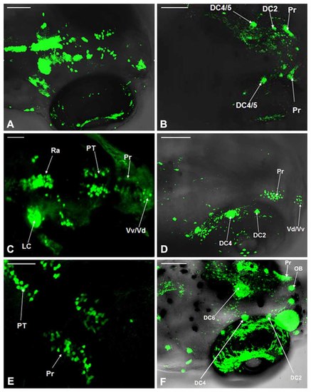
Comparison of dopaminergic neuron distribution in the Vmat2:GFP and WT zebrafish. The distribution of dopaminergic neurons in WT zebrafish at 4 dpf (A), 5 dpf (C) and 6 dpf (E) was detected using TH whole mount immunofluorescent and compared with that in Vmat2:GFP transgenic zebrafish at 4 dpf (B), 5 dpf (D) and 6 dpf (F) correspondingly. DC, diencephalons; Pr, pretectum; PT, posterior tubercle; LC, locus coeruleus; Vd/Vv, dorsal/ventral nucleus of the telencephalic area; Ra, raphe nuclei; OB, olfactory bulb. Scale: (A,B,D,F) = 100 μm; (C,E) = 50 μm. |
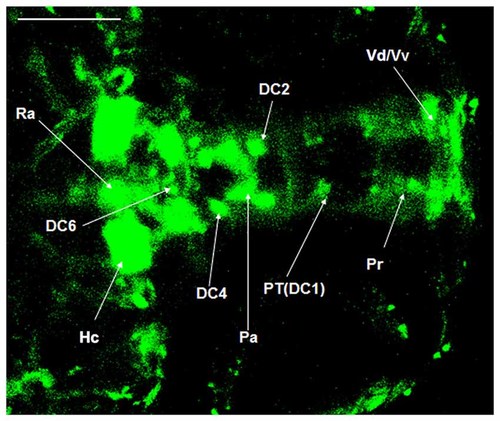
Localization of dopaminergic nuclei in Vmat2:GFP zebrafish. The dopaminergic nuclei in the ventral DC of the Vmat2:GFP zebrafish at 4 dpf were identified under laser scanning confocal microscope (LSCM). All the dopaminergic neurons had already been developed. DC, diencephalons; Pa, paraventricular nucleus; Pr, pretectum; PT, posterior tubercle; Vd/Vv, dorsal/ventral nucleus of the telencephalic area; Ra, raphe nuclei; Hc, caudal hypothalamus. Scale = 100 μm. |
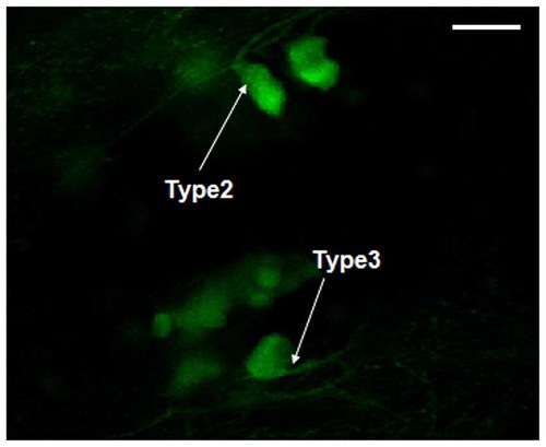
Morphology observation of dopaminergic neurons in Vmat2:GFP zebrafish. According to the classification of dopaminergic neurons (Kawakami et al., 2000), the observed dopaminergic neurons were mainly Type 2 and Type 3. Scale = 50 μm. |

Axonal projections of dopaminergic neurons in Vmat2:GFP zebrafish. The axonal projections from periventricular nucleus of posterior tubercle (TPp) to subpallium (A,B) were found at 5 dpf. TPp, periventricular nucleus of posterior tubercle. Scale = 40 μm. |