
FIGURE
Fig. 4
- ID
- ZDB-FIG-170130-11
- Publication
- Du et al., 2016 - Spatial and Temporal Distribution of Dopaminergic Neurons during Development in Zebrafish
- Other Figures
- All Figure Page
- Back to All Figure Page
Fig. 4
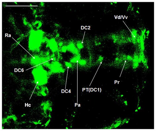
Localization of dopaminergic nuclei in Vmat2:GFP zebrafish. The dopaminergic nuclei in the ventral DC of the Vmat2:GFP zebrafish at 4 dpf were identified under laser scanning confocal microscope (LSCM). All the dopaminergic neurons had already been developed. DC, diencephalons; Pa, paraventricular nucleus; Pr, pretectum; PT, posterior tubercle; Vd/Vv, dorsal/ventral nucleus of the telencephalic area; Ra, raphe nuclei; Hc, caudal hypothalamus. Scale = 100 μm. |
Expression Data
Expression Detail
Antibody Labeling
Phenotype Data
Phenotype Detail
Acknowledgments
This image is the copyrighted work of the attributed author or publisher, and
ZFIN has permission only to display this image to its users.
Additional permissions should be obtained from the applicable author or publisher of the image.
Full text @ Front. Neuroanat.