
Fig. 5
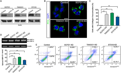
SCFD1 and STX18 are necessary for Procollagen II transport. (A) Efficiency of knockdown of SCFD1, TANGO1 and STX18 as measured by western blotting. (B) Quantification of cells that accumulate Procollagen II intracellularly. The percentage of cells that accumulate Procollagen II intracellularly was determined by counting at least 30 cells in five random fields. The number of cells accumulating Procollagen II in the case of SCFD1, TANGO1 and STX18 shRNA was significantly elevated as compared to control shRNA cells. Error bars indicate s.e.m. **P<0.01 as determined by Student's t-tests. (C) ATDC5 cells were visualized with anti-Collagen II antibody and DAPI. Scale bar, 20 µm. (D) Elevated XBP1 mRNA splicing in ATCD5 cell was detected by RT-PCR and Q-PCR. XBP1-u, unspliced XBP1 mRNA. XBP1-s, spliced XBP1 mRNA. β-actin served as a loading control. Error bars represent mean±SEM. **P<0.01 as determined by Student's t-tests. (E) Apoptosis in ATDC5 cells detected by FACS analysis. The right quadrants (Q2 and Q3) represent apoptotic cells, with cytoplasmic membrane integrity. The average apoptotic rates of control, SCFD1 knockdown, TANGO1 knockdown and STX18 knockdown samples were 2.51±0.56, 35.11±0.40, 36.43±1.31 and 28.58±2.32%, respectively. |
Reprinted from Developmental Biology, 421(1), Hou, N., Yang, Y., Scott, I.C., Lou, X., The Sec domain protein Scfd1 facilitates trafficking of ECM components during chondrogenesis, 8-15, Copyright (2017) with permission from Elsevier. Full text @ Dev. Biol.