
Fig. 3
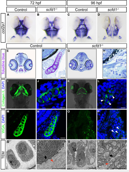
Protein trafficking is disrupted in scfd1 mutants. (A–D) Chondrocytes in scfd1 mutant embryo express col2a1a normally at 72 hpf, with reduced expression by 96 hpf. A to D, ventral views with anterior to the top. (E-H) Toluidine blue staining of transverse sections of the jaw at 80 hpf. Nuclei are stained blue, whereas ECM is stained purple. (I–P) Immunostaining of Collagen II and WGA in the palatoquadrate of 80 hpf wild type and scfd1 mutant embryos. Arrowheads in L and P indicate aberrant intracellular ECM protein localization in a scfd1 mutant. (Q–T) TEM images of 80 hpf wild type and scfd1 mutant chondrocytes. Black arrows indicate well-stacked ER in wild type cell (R) or distended ER membranes in scfd1 mutant cells (T). Orange arrowheads point to collagen fibril deposition in the extracellular space. Scale bars: 25 µm (F, H, J, L, N and P); 2 µm (Q–T). For A to P, at least 15 embryos for each genotype were analyzed and representative samples are shown. For Q to T, 6 embryos for each genotype were analyzed and representative samples are shown. |
| Gene: | |
|---|---|
| Antibody: | |
| Fish: | |
| Anatomical Terms: | |
| Stage Range: | Protruding-mouth to Day 4 |
| Fish: | |
|---|---|
| Condition: | |
| Observed In: | |
| Stage Range: | Protruding-mouth to Day 4 |
Reprinted from Developmental Biology, 421(1), Hou, N., Yang, Y., Scott, I.C., Lou, X., The Sec domain protein Scfd1 facilitates trafficking of ECM components during chondrogenesis, 8-15, Copyright (2017) with permission from Elsevier. Full text @ Dev. Biol.