
Fig. 2
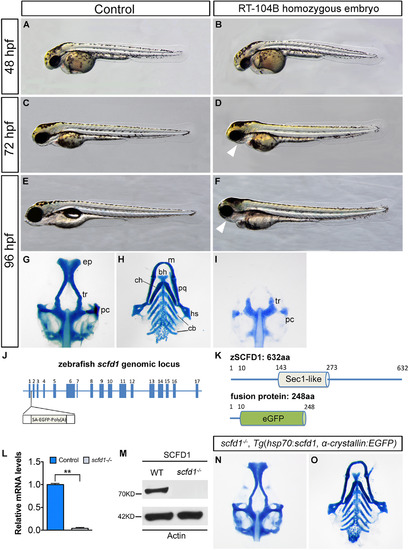
Loss of scfd1 disrupts craniofacial skeletal development. (A–F) Live images of wild type (control) and RP24-104B homozygous embryos at indicated developmental stages. Lateral view with head to left. (G–I, N and O) Alcian blue staining of cartilage elements in head skeleton in ventral views. White arrowheads indicate dismorphic jaw. bh, basihyal; cb, ceratobranchial; ch, ceratohyal; ep, ethmoid plate; hs, hyosymplectic; m, Meckel′s cartilage; pc, parachordal plate; pq, palatoquadrate; tr, trabeculae. (J) The zebrafish scfd1 genomic locus. The transposon is inserted after the first exon. (K) Schematic representations of the domain structure of the wild type zebrafish SCFD1 protein and fusion protein in the gene trap scfd1 mutant. (L and M) Q-PCR and western blotting experiments confirmed loss of scfd1 expression in homozygous mutant embryos at 48 hpf. **P<0.01 as determined by Student's t-tests. Error bars indicate s.e.m. (N and O) Rescue of the scfd1 mutant phenotype in Tg(hsp70:scfd1; α-crystallin:EGFP) embryos at 96 hpf. Heat shock was performed every 12 h from 24 hpf onwards. |
| Fish: | |
|---|---|
| Condition: | |
| Observed In: | |
| Stage Range: | Long-pec to Days 7-13 |
Reprinted from Developmental Biology, 421(1), Hou, N., Yang, Y., Scott, I.C., Lou, X., The Sec domain protein Scfd1 facilitates trafficking of ECM components during chondrogenesis, 8-15, Copyright (2017) with permission from Elsevier. Full text @ Dev. Biol.