
Fig. 2
- ID
- ZDB-FIG-161207-6
- Publication
- Hamilton et al., 2016 - A Zebrafish Live Imaging Model Reveals Differential Responses of Microglia Toward Glioblastoma Cells In Vivo
- Other Figures
- All Figure Page
- Back to All Figure Page
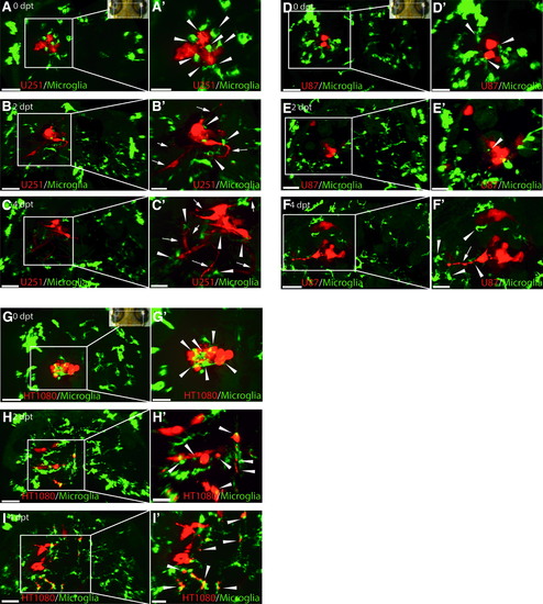
U251, U87, and HT1080 cells exhibit different growth patterns in the zebrafish optic tectum and stimulate an intensive microglia response. Representative confocal images of the optic tectum of mpeg1:EGFP zebrafish transplanted with U251mCherry (U251) glioblastoma cells, U87mCherry (U87) glioblastoma cells, and HT1080mCherry (HT1080) fibrosarcoma cells at 3 dpf. (A–C) Images from top to bottom are in chronological order showing 0, 2, and 4 dpt of U251 cells; (A′–C′) Higher magnification of regions of interest in (A–C) to highlight interactions of microglia (green) and U251 cells (red). U251 cellular projections are marked with a white arrow. Microglia interacting with U251 cells are marked with a white arrowhead. (D–F) Images from top to bottom are in chronological order showing 0, 2, and 4 dpt of U87 cells; (D′–F′) Higher magnification of regions of interest in (D–F) to highlight interactions of microglia (green) and U87 cells (red). U87 cellular projections are marked with a white arrow. Microglia interacting with U87 cells are marked with a white arrowhead. (G–I) Images from top to bottom are in chronological order showing 0, 2, and 4 dpt of HT1080 cells; (G′–I′) Higher magnification of regions of interest (G–I) to highlight interactions of microglia (green) and HT1080 cells (red). Microglia interacting with HT1080 cells are marked with a white arrowhead. Scale bars for (A–I): 50 μm. Scale bars for (A′–I′): 30 μm. All images represent maximum intensity projections of confocal stacks. Images were captured using an Andor spinning disk confocal microscope with a 20 × /NA 0.75 objective. Insets in (A, G, and D) show the orientation of samples (anterior to the top) and the region imaged (white rectangle). dpf, days postfertilization; dpt, days posttransplantation |
| Gene: | |
|---|---|
| Fish: | |
| Condition: | |
| Anatomical Term: | |
| Stage: | Protruding-mouth |