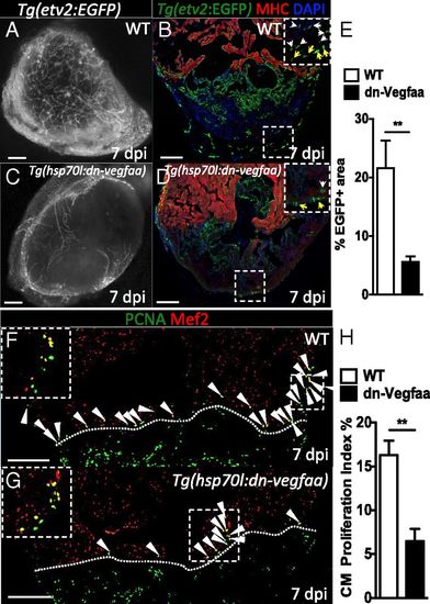
Revascularization of the damaged area supports CM proliferation. (A and C) Images of the apex of Tg(etv2:EGFP) WT and Tg(hsp70l:dn-vegfaa);Tg(etv2:EGFP) ventricles at 7 dpi. WT and Tg(hsp70l:dn-vegfaa) were daily heatshocked. (B and D) Sections of Tg(etv2:EGFP) WT and Tg(hsp70l:dn-vegfaa);Tg(etv2:EGFP) ventricles. CMs are immunostained with anti-MHC antibody (red), and nuclei counterstained with DAPI (blue). Insets show high-magnification images of the coronaries on the surface of the heart (yellow arrows) and in the damaged area (white arrows). (E) Quantification of coronary vessel formation in the injured area. The percentage of etv2:EGFP+ area was measured relative to total injured area in WT ventricles (n = 6) and Tg(hsp70l:dn-vegfaa) ventricles (n = 5). (F and G) Sections from Tg(etv2:EGFP) WT and Tg(hsp70l:dn-vegfaa);Tg(etv2:EGFP) ventricles stained with anti-Mef2 (red) and anti-PCNA (green) antibodies. Arrowheads point to Mef2+/PCNA+ nuclei. Insets show high-magnification images of Mef2+/PCNA+ nuclei. (H) Quantification of CM proliferation in WT (n = 5) and Tg(hsp70l:dn-vegfaa) (n = 5) ventricles at 7 dpi. Data are expressed as mean ± SEM, **P < 0.001. Dotted lines delineate the injured area. (Scale bars: 100 µm.)
|
