
FIGURE
Fig. S2
- ID
- ZDB-FIG-161103-14
- Publication
- Sicca et al., 2016 - Gain-of-function defects of astrocytic Kir4.1 channels in children with autism spectrum disorders and epilepsy
- Other Figures
- All Figure Page
- Back to All Figure Page
Fig. S2
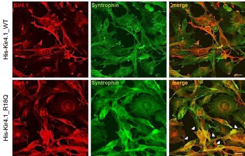
Double immunofluorescence stainings with anti-Kir4.1 pAb (red) and anti-synt mAb (green) in U251 cells expressing WT (upper panels) or R18Q (lower panels) Kir4.1 reveal a partial colocalization of Kir4.1 and syntrophin in the plasma membrane and in the cytoplasm of both astrocytoma cell lines. Compared to Kir4.1 WT expressing cells, a larger number of R18Q+ cells shows colocalization of syntrophin and Kir4.1 in both cytoplasm and plasma membrane (arrowheads). Scale bars: 10 µm. |
Expression Data
Expression Detail
Antibody Labeling
Phenotype Data
Phenotype Detail
Acknowledgments
This image is the copyrighted work of the attributed author or publisher, and
ZFIN has permission only to display this image to its users.
Additional permissions should be obtained from the applicable author or publisher of the image.
Full text @ Sci. Rep.