Fig. 5
|
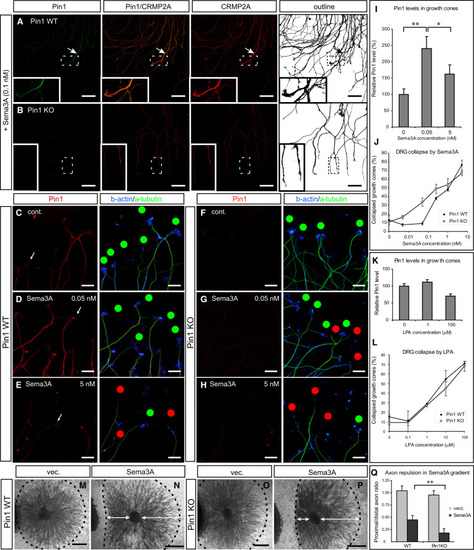
Pin1 KO Increases Sensitivity to Sema3A-Induced Growth Cone Collapse in Primary Dorsal Root Ganglia Neurons (A and B) Sema3A induces colocalization of high levels of Pin1 and CRMP2A in the vicinity of growth cones (arrows) in Pin1 WT, but not Pin1 KO, DRG axons. Pin1 WT (A) and KO (B) primary DRG neurons were treated with 0.1 nM Sema3A for 30 min, followed by double immunostaining for Pin1 (green) and CRMP2A (red). (C-J) Pin1 KO increases sensitivity to Sema3A-induced growth cone collapse. Pin1 WT (C-E) and KO (F-H) primary DRG neurons were treated with different concentrations of Sema3A for 30 min, fixed, and triple immunostained with anti Pin1, β-actin, and β-tubulin antibodies, followed by growth cone collapse analysis, with the percentage of growth cone collapse being shown in (J). Red dots, collapsed growth cones; green dots, intact growth cones. Intensity of Pin1 immunostaining in DRG growth cones (C) significantly increases upon low (non-collapsing) Sema3A stimulation (D) and is reduced upon high Sema3A stimulation (E); the quantification after normalization to ²-tubulin levels is shown (I). (K and L) Stimulation with LPA induces similar growth cone collapse in Pin1 WT and KO DRG neurons (L) and does not affect Pin1 levels in the growth cones (K). (M-Q) Pin1 KO significantly increases sensitivity to Sema3A-induced growth cone collapse in collagen 3D co-cultures. SH-SY5Y cells were transfected with empty vector (M and O) or Sema3A expression vector (N and P) and co-cultured with Pin1 WT (M and N) or KO (O and P) DRG. Proximal/distal axon length ratio was measured (N and P, arrows) upon NF-M immunostaining and quantified (Q). Scale bars represent 50 µm (A and B), 20 µm (C-H), and 500 µm (M-P); *p < 0.05; **p < 0.0001. Values are means ± SEM. See also Figure S4. |

