
FIGURE
Fig. S8
- ID
- ZDB-FIG-160826-24
- Publication
- Ji et al., 2016 - Mutations in zebrafish pitx2 model congenital malformations in Axenfeld-Rieger syndrome but do not disrupt left-right placement of visceral organs
- Other Figures
- All Figure Page
- Back to All Figure Page
Fig. S8
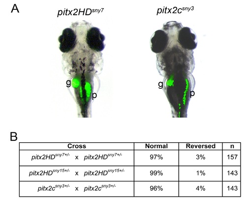
Normal asymmetric placement of visceral organs in pitx2 mutants. (A) Representative images of auto-fluorescence in the gall bladder (g) and pancreas (p) of 7 dpf larvae. L=left; R=right. (B) Scoring of organ placement in larvae from pitx2 heterozygous parents (25% expected to be homozygous mutants). n=number of embryos analyzed. |
Expression Data
Expression Detail
Antibody Labeling
Phenotype Data
Phenotype Detail
Acknowledgments
This image is the copyrighted work of the attributed author or publisher, and
ZFIN has permission only to display this image to its users.
Additional permissions should be obtained from the applicable author or publisher of the image.
Reprinted from Developmental Biology, 416(1), Ji, Y., Buel, S.M., Amack, J.D., Mutations in zebrafish pitx2 model congenital malformations in Axenfeld-Rieger syndrome but do not disrupt left-right placement of visceral organs, 69-81, Copyright (2016) with permission from Elsevier. Full text @ Dev. Biol.