
Fig. 4
- ID
- ZDB-FIG-160726-9
- Publication
- Hensley et al., 2016 - Evolutionary and developmental analysis reveals KANK genes were co-opted for vertebrate vascular development
- Other Figures
- All Figure Page
- Back to All Figure Page
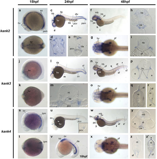
Kank2, kank3 and kank4 gene expression during zebrafish development. Whole mount in situ hybridization of zebrafish embryos at stages 15 hpf (a,b,j,k,s,t), 18 hpf (v), 24 hpf (c-e,l,m,u) and 48 hpf (f-i,n-q,w-z′). Anterior is to the left in all whole-mount images, and dorsal is to the top in all transverse sections. (a-i). Gene expression of kank2. (j-r). Gene expression of kank3. (s-z′). Gene expression of kank4. The dashed lines indicate the positions of section. The letters below the dashed lines correspond to the panels. The inset within panel (u) is the transverse section of the dashed line above. The white arrow points to the kank4 expression on the vascular vessels on the surface of the yolk. af, anal fin bud; cf, caudal fin bud; cv, caudal vein; da, dorsal aorta; df, dorsal fin bud; dlv, dorsal longitudinal vein; e, eye; hb, hindbrain; hv, head vessels; llp, lateral line premordia; psm, presomatic mesoderm; lpm, lateral plate mesoderm; mb, midbrain; nt, neural tube; ov, otic vesicle; pa, pharyngeal arch; pcv, posterior cardinal vein; pf, pectoral fin bud; so, somite. |
| Genes: | |
|---|---|
| Fish: | |
| Anatomical Terms: | |
| Stage Range: | 10-13 somites to Long-pec |